Indocyanine green angiography, or ICG angiography for short, is used primarily in vascular neurosurgery. This simple and harmless procedure can be used to visualize the blood flow through the cerebral vessels in real time during surgery. This allows the surgeon to monitor blood flow to the vessels while the procedure is still in progress. ICG angiography thus improves safety and treatment outcomes in vascular surgery for aneurysms or arteriovenous malformations.
How does ICG angiography work?
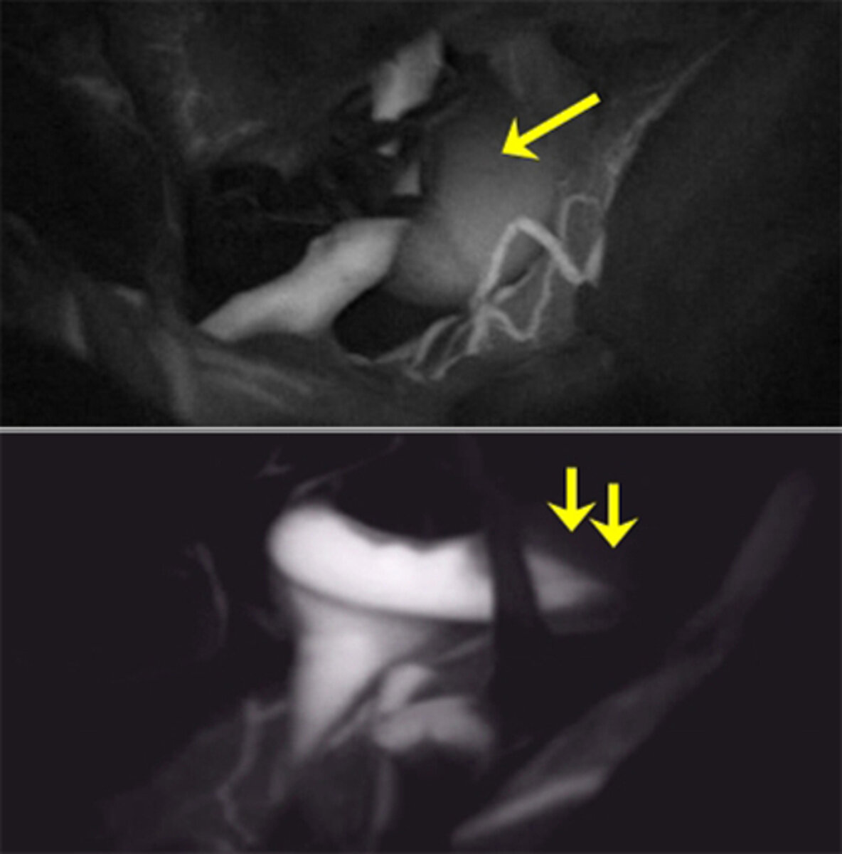
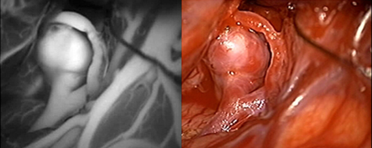
Indocyanine green (ICG) is a liquid fluorescent dye that is harmless to humans. During surgery, a few milliliters of ICG are administered through venous access and rapidly pumped into the arterial bloodstream. Simultaneously, invisible light with a wavelength of approximately 800 nm is directed onto the brain surface from the surgical microscope specially equipped for this purpose *,*. As soon as the ICG reaches the brain vessels together with the arterial blood flow, the vessels glow white. After passing through the cerebral arteries, the brain tissue and consequently the draining cerebral veins contrast.
This not only provides the surgeon with information about whether a vessel is open or occluded, but also allows them to distinguish veins from arteries and assess the dynamics of flow. A video of the flow is created, which can be viewed several times by the surgeon or used for additional flow analysis.
ICG angiography only takes a few seconds. The fluorescent dye remains in the vascular system for approximately 20 minutes and is then completely excreted from the body.
How is ICG angiography used at Inselspital?
We use ICG angiography primarily for vascular neurosurgery procedures – especially for the microsurgical treatment of aneurysms and arteriovenous malformations.
Aneurysm surgery
ICG angiography offers two advantages here:
- Using this technique, the surgeon can immediately assess the patency of the unaffected surrounding vessels after applying the clip. An incorrectly positioned clip, which occludes not only the aneurysm but also outgoing vessels, can thus be identified and repositioned immediately. This may prevent a subsequent stroke.
- In addition, the surgeon can immediately check whether the aneurysm has been completely occluded. If the position of the clip is not optimal, it can still be repositioned.
In an international, prospective, controlled study with more than 100 patients, ICG angiography showed an intraoperative finding in 9% of operations, which led to a change in management and repositioning of a clip *. This clearly demonstrates the importance of ICG angiography for the safety of aneurysm surgery and how much it improves closure rates after clipping.
AVM surgery
The use of ICG angiography has also shown clear advantages in arteriovenous malformation (AVM) surgery. It can be used during the procedure to differentiate between feeding and draining vessels, analyze the flow in the vascular tumor and verify complete resection at the end of the operation *.
Our experience at Inselspital
ICG angiography was introduced to neurosurgery by our director and chief physician Prof. Andreas Raabe *,*,* and further developed together with Prof. Robert Spetzler from Phoenix in Arizona, USA. The procedure is now standard in vascular neurosurgery at Inselspital – especially in the operation of aneurysms and arteriovenous malformations.
-
Raabe A, Beck J, Gerlach R, Zimmermann M, Seifert V. Near-infrared indocyanine green video angiography: a new method for intraoperative assessment of vascular flow. Neurosurgery. 2003 Jan;52(1):132-9; discussion 139.
-
Raabe A, Beck J, Seifert V. Technique and image quality of intraoperative indocyanine green angiography during aneurysm surgery using surgical microscope integrated near-infrared video technology. Zentralbl Neurochir. 2005 Feb;66(1):1-6; discussion 7-8.
-
Raabe A, Nakaji P, Beck J, Kim LJ, Hsu FPK, Kamerman JD, et al. Prospective evaluation of surgical microscope-integrated intraoperative near-infrared indocyanine green videoangiography during aneurysm surgery. J Neurosurg. 2005 Dec;103(6):982-9.
-
Killory BD, Nakaji P, Gonzales LF, Ponce FA, Wait SD, Spetzler RF. Prospective evaluation of surgical microscope-integrated intraoperative near-infrared indocyanine green angiography during cerebral arteriovenous malformation surgery. Neurosurgery. 2009 Sep;65(3):456-62; discussion 462.