Augmented reality is a digital technology that supplements reality with additional virtual information. These virtual extensions can be texts, graphics, animations, videos, static or moving 3-D objects. The user can perceive the virtual elements in his real environment with the help of special AR glasses. This technology is primarily known in the gaming sector, but is increasingly being used in other areas as well. In medicine, for example.
Making the invisible visible
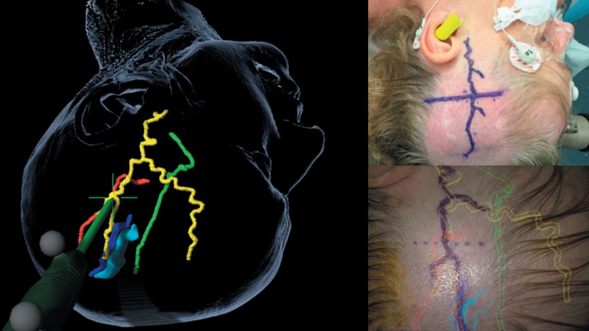
Structures, tumors and functional centers lie hidden in the depths of the brain and are not visible to the naked eye. The same applies to nerves or blood vessels that run embedded and hidden in the tissue. Using very high-resolution images from magnetic resonance imaging (MRI) or computed tomography (CT), the location of a tumor or the course of vessels or nerves can be displayed in a 3D model.
If this 3D model is projected exactly onto the patient's head, this data can be superimposed on the surgical microscope. When looking at the patient, the surgeon then sees not only the real image but also the superimposition of invisible structures. This is why we also speak of mixed reality, i.e. a mixture of reality (the patient) and image (the tumor or the vessel).
This procedure is extremely helpful for the surgeon, as he is precisely informed about the position of important structures of the brain or the spine during the operation. This can increase accuracy during surgery, which in turn means greater safety for the patient and improves the surgical outcome.
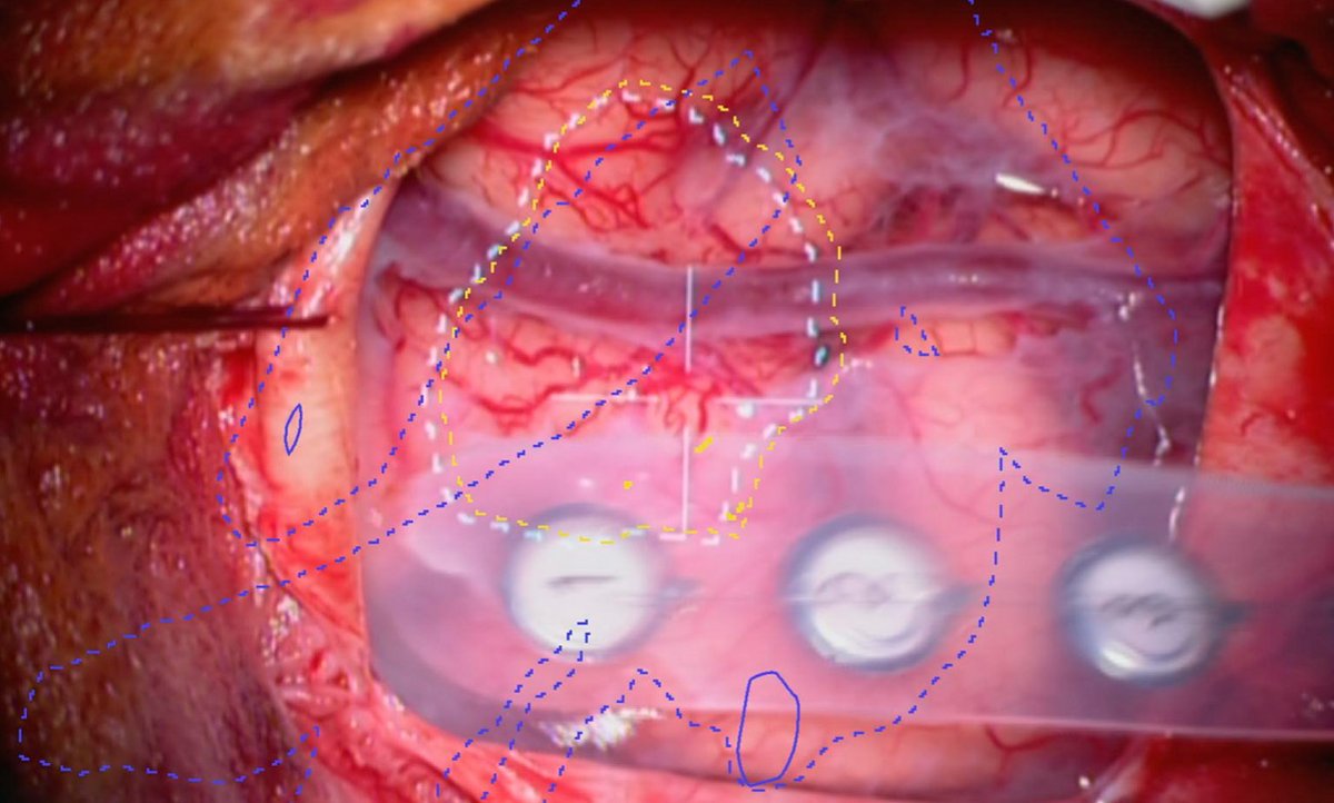
Cerebral vascular surgery
One area of application for augmented reality is vascular navigation in cerebrovascular surgery. This makes it easier to locate vessels during surgery and to operate on vascular malformations or diseases in a more targeted manner.
Tumor surgery
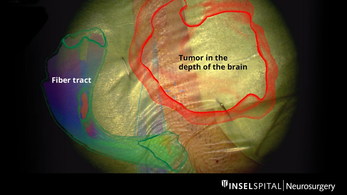
As a general rule, neurosurgery should remove as much tumor tissue as possible. The tumor should be radically removed up to the border of the functioning brain tissue. However, since it is often difficult to distinguish between tumor and healthy brain tissue, especially in the marginal area, augmented reality is also used here – in addition to other modern technologies.
The powerful surgical microscope allows fine work on the brain with millimeter precision. The fiber tracts already drawn in before the operation, as well as the tumor itself and other important centers, are virtually superimposed in the surgical microscope and projected onto the surface of the head. This enables the surgeon to orientate himself better and to plan the access to the tumor in a tailor-made way: as small as possible, but as large as necessary.
How do we develop AR further?
Augmented reality is a research focus of our clinic. We currently use augmented reality on a daily basis during routine operations by superimposing structures in the eyepiece of the surgical microscope. Currently, we are researching the improvement of this procedure. The goal is to detach augmented reality from the surgical microscope. With special AR glasses, it would be possible to use the technology without a microscope. This would be helpful in spinal surgeries, where the surgeon could capture the herniated disc or a narrowing in the spinal canal with millimeter precision and apply the smallest possible approach. We see great potential in this technology for the future