Positron emission tomography, or PET for short, is an imaging technique from nuclear medicine, with which metabolic activities in tissue can be depicted visually. It is primarily used for detecting cancer and its metastases.
In PET, a molecule is administered to the patient that allows the visualization of particularly active tumor areas. We usually use fluoroethyltyrosine (FET) for this purpose. FET is a weakly radioactively labeled amino acid and indicates the increased amino acid metabolism in tumor cells. Pathological changes can thus be effectively depicted visually.
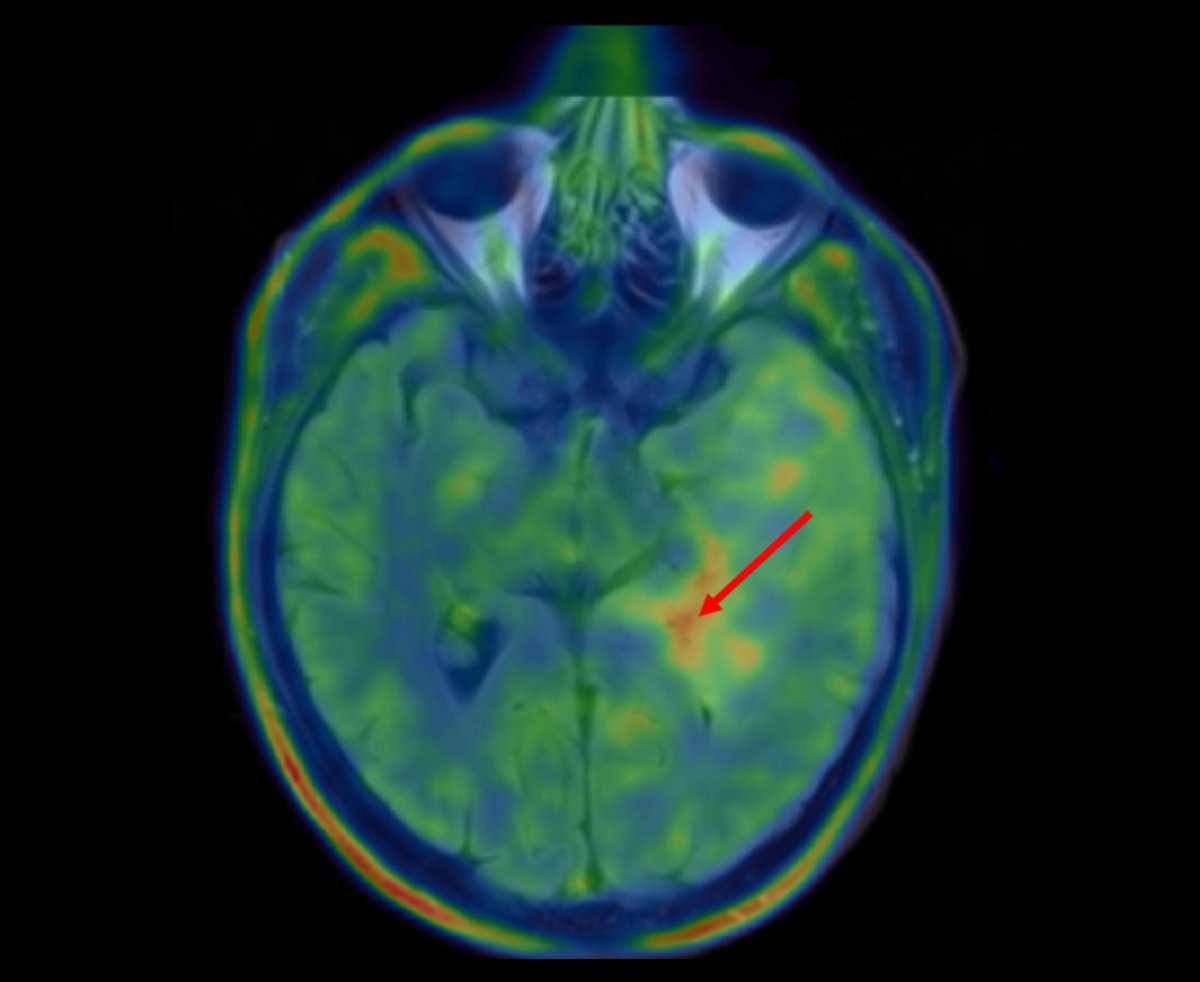
By overlaying PET data with MRI data, a hotspot with particularly active tumor cells can often be identified. This hotspot represents a good target for a biopsy.
At the end of 2020, the world's fastest PET-CT scanner was put into operation at the University Department of Nuclear Medicine at Inselspital. This scanner of the very latest technology enables excellent examination quality with shorter examination times and reduced radiation exposure for our patients.