A Rathke cleft cyst is a benign cyst in the brain. It is a remnant from the embryonic development of the pituitary gland (hypophysis) that has not regressed. If the Rathke cleft cyst is small and does not cause symptoms, it does not need therapy. However, if it puts pressure on adjacent brain structures, symptoms may occur. On this page you will learn what symptoms a Rathke cleft cyst can cause and how we can help you at Inselspital to treat it.
How does a Rathke cleft cyst develop?
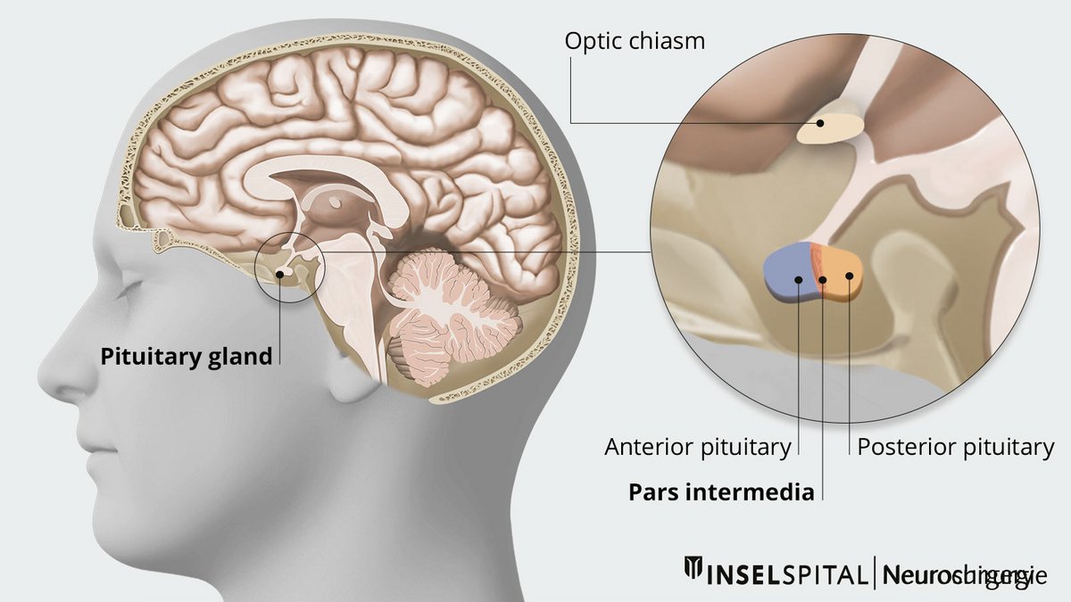
Rathke cleft cyst is a malformation of remnants of Rathke pouch that normally regresses completely during embryonic development of the pituitary gland. Rathke cleft cysts are congenital and usually originate in the middle part of the pituitary gland. They may increase in size and expand in various directions during life.
How common is a Rathke cleft cyst?
Rathke cleft cysts are not that rare and are often discovered by chance. The prevalence in the population is about 13-23%. Women are more frequently affected than men, approximately in a ratio of 2:1.
What are the symptoms of a Rathe cleft cyst?
Most Rathke cleft cysts are small and asymptomatic. If they grow and exert pressure on adjacent brain areas, the following symptoms may occur:
- headache
- visual disturbances and/or visual field restrictions due to compression of the optic nerves
- hormonal disturbances due to compression of the pituitary gland
How is a Rathke cleft cyst diagnosed?
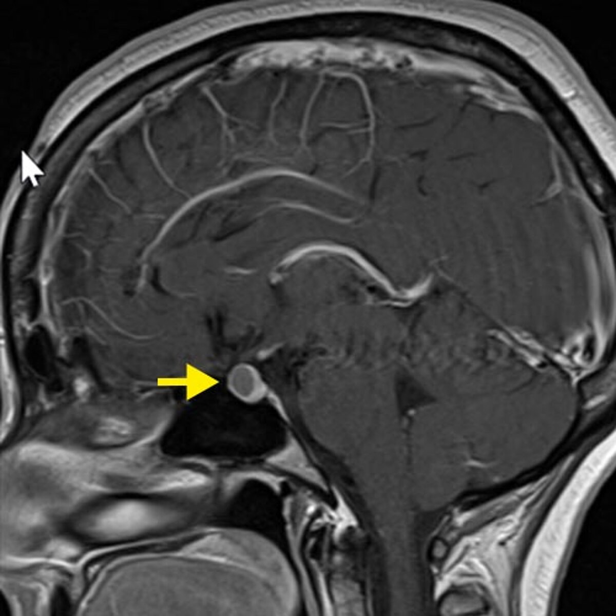
A magnetic resonance imaging (MRI) is the diagnostic procedure of first choice. The MRI image provides information on the localization and extent of the cyst.
In patients with a larger cyst, an endocrinologic workup is advisable to diagnose and treat possible hormonal imbalances.
If the MRI result shows that the cyst is in contact with the optic chiasm, the place where the optic nerves cross, an ophthalmological examination is also organized. The ophthalmologist can detect and quantify visual acuity and visual field loss. These usually remain unnoticed by the affected person for a long time.
How is a Rathke cleft cyst treated?
In small asymptomatic Rathke cleft cysts, we arrange for follow-up by MRI at intervals of 6-12 months to exclude an increase in size. Very small cysts (< 5 mm) do not require monitoring.
When a Rathke cleft cyst causes discomfort, surgical therapy is indicated. The operation is usually performed via a transnasal transsphenoidal approach. The procedure can be performed microsurgically, endoscopically assisted or fully endoscopically at the Inselspital. Since the cyst wall is often strongly attached to surrounding structures such as the pituitary stalk or optic chiasm, partial resection or fenestration of the cyst is usually performed.
Possible complications after resection of a Rathke cleft cyst are primarily disturbances of the electrolyte and hormone balance. These disturbances are usually temporary. In the postoperative phase, blood values and water balance are monitored closely. If necessary, hormones must be substituted with medication.
Rathke cleft cysts recur not at all infrequently, so that a new operation may become necessary in the course of time.
Why you should seek treatment at Inselspital
- Our specialized neurosurgical team has many years of experience in the treatment of Rathke cleft cysts.
- We work closely with the endocrinology and ophthalmology departments to provide comprehensive, high-quality care for our patients before, during and after surgery. The best possible treatment strategy is determined for each individual patient. This is done within the framework of our interdisciplinary pituitary board, where the individual therapy plan is developed.
- For surgery we use innovative technical procedures such as neuronavigation and intraoperative neuromonitoring. These new achievements are a guarantee for maximum precision during surgery and maximum safety for our patients.
Further reading
- Chotai S, Liu Y, Pan J, Qi S. Characteristics of Rathke's cleft cyst based on cyst location with a primary focus on recurrence after resection. J Neurosurg. 2015 Jun;122(6):1380-9.
- Greenberg MS. Handbuch der Neurochirurgie. 8. Aufl. Thieme 2016.
- Zada G. Rathke cleft cysts: a review of clinical and surgical management. Neurosurg Focus.2011 Jul;31(1):E1.